
Dr. Gaurav Verma is a postdoctoral fellow at the Icahn School of Medicine at Mount Sinai specializing in the development of accelerated and highly-sensitive spectroscopy and imaging sequences for ultra-high field MRI. He has implemented these sequences in the study of human gliomas, psychiatric disorders such as major depression and schizophrenia, and neurological disorders including amyotrophic lateral sclerosis, multiple sclerosis and epilepsy. Dr. Verma’s ongoing work explores novel imaging sequences to characterize highly sensitive imaging and metabolic biomarkers for neuropsychiatric disorders, and image processing techniques employing conventional & machine learning based strategies to glean additional data from the acquired images.
Peer-Reviewed Publications
- Verma G, et al. Whole Brain Echo-planar Spectroscopic Imaging for Differentiation of Trie Progression from Pseudoprogression in Patients with Glioblastoma, NMR in Biomedicine 2018 In Revision
- Chawla S et al. Differentiation of Brain Infection from Necrotic Glioblastoma using Combined Analysis of Diffusion and Perfusion Magnetic Resonance Imaging JMRI 2018 DOI: 10.1002/jmri.26053
- Bagga P, et al. In vivo GluCEST MRI: Reproducibility, Background contribution and source of glutamate changes in the MPTP model of Parkinson’s disease, Scientific Reports 2018 DOI:10.1038/s41598-018-21035-3
- Gurbani S, et al. Convolutional Neural Network to Filter Artifacts in Spectroscopic Magnetic Resonance Imaging Magn Reson Med. 2018 DOI: 10.1002/mrm.27166
- Verma G, et al. Average weighted acquisition for faster localized two-dimensional correlated spectroscopy of the brain in vivo. JMR 2017; 227:104-112
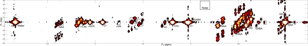
- Verma G, et al. Non-invasive detection of 2-hydroxyglutarate in IDH-mutated gliomas using two-dimensional localized correlation spectroscopy (2D L-COSY) at 7 Tesla. Journal of Translational Medicine 2016 14:274
- Chawla S et al. Dynamic Contrast-Enhanced MRI-Derived Intracellular Water Lifetime (τ i ): A Prognostic Marker for Patients with Head and Neck Squamous Cell Carcinomas. AJNR 2018 39:138-144
- Bagga P, et al. Mapping the alterations in glutamate with GluCEST MRI in a mouse model of dopamine deficiency. Journal of Neurochemistry 2016; 139:432-439
- Mohan S et al. Assessment of early response to tumor-treating fields in newly diagnosed glioblastoma using physiologic and metabolic MRI: initial experience. CNS Oncology 2016 Vol. 5, No. 3
- Kumar M, et al. Magnetic Resonance Spectroscopy for Detection of Choline Kinase Inhibition in the Treatment of Brain Tumors, Mol Cancer Ther 2015; 14:899
- Verma G, et al. Implementation of two‐dimensional L‐COSY at 7 tesla: An investigation of reproducibility in human brain. J. Magn. Reson. Imaging 2014; 40:1319-1327
- Haris M, et al. High Resolution Mapping of Modafinil Induced Changes in Glutamate Level in Rat Brain. 2014 PLOS ONE. 9(7): e103154
- Verma G, et al. Whole-Brain Analysis of Amyotrophic Lateral Sclerosis by Using Echo-Planar Spectroscopic Imaging. Radiology 2013; 267(3), 851-857.
- Sahin N, et al. Advanced MR Imaging Techniques in the Evaluation of Nonenhancing Gliomas: Perfusion-Weighted Imaging Compared with Proton Magnetic Resonance Spectroscopy and Tumor Grade. Neuroradiology Journal 2013; 26(5).
- Haris M, et al. Imaging of glutamate neurotransmitter alterations in Alzheimer’s disease. NMR in Biomed 2013; 26(4):386-391
- Nagarajan R, et al. MR spectroscopic imaging and diffusion-weighted imaging of prostate cancer with Gleason scores. J. Magn. Reson. Imaging 2012; 36: 697–703.
- Verma G, et al. Implementation of Multi-Echo Based Correlated Spectroscopic Imaging and Preliminary Findings in Human Brain and Calf Muscle. JMRI. 2011; 34:262-269.
- Lipnick S, et al. Echo planar correlated spectroscopic imaging: Implementation and pilot evaluation in human calf in vivo. Magn Reson Med. 2010; 64:947-956.
Recent Conference Abstracts
- Verma G, et al. Assessment of metabolic heterogeneity in glioblastoma multiforme (GBM) through histogram analysis of whole-brain echo planar spectroscopic imaging. ISMRM 2018
- Verma G, et al. Spectroscopic Imaging-based detection of 2-hydroxyglutarate (2HG) in IDH1 mutant human gliomas on 3T Clinical ISMRM 2017
- Verma G, et al. Whole-brain echo planar spectroscopic imaging distinguishes recurrent tumor versus pseudoprogression in glioblastoma patients. ISMRM 2016
- Verma G, et al. Development and Implementation of a Matlab-based multi-modal 3D visualization, co-registration and quantification platform for assessing brain tumor physiology and metabolism ISMRM 2016
- Verma G, et al. Chemical Shift Imaging (CSI) for detection of 2-hydroxyglutarate (2HG) in human gliomas at 3T RSNA 2015
- Verma G, et al. Two-dimensional localized correlated spectroscopy (2D L-COSY) at 7T for detection of 2-hydroxyglutarate in gliomas with IDH mutations. RSNA 2015
- Verma G, et al. Average weighted acquisition for faster acquisition of in vivo localized two dimensional correlation spectroscopy of the brain. ISMRM 2015
- Verma G, et al. 3D Echo-Planar Spectroscopic Imaging detects Pseudoprogression in Glioblastoma Patients. ISMRM Cancer Workshop 2014
- Verma G, et al. Single voxel two dimensional correlation spectroscopy of brain tumors at 7T. ISMRM Cancer Workshop 2014
- Verma G, et al. 2D L-COSY at 7T Detects Glutamate, Glutamine, Glutathione and GABA in Patients with Schizophrenia. ISMRM 2014
- Verma G, et al. A MATLAB-Based Program for Post-Processing Across Multiple Platforms. ISMRM 2014
- Verma G, et al. Case Study: 2D L-COSY of Low-Grade Gliomas at 7T. ISMRM 2014